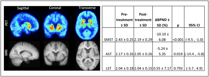
Figure 1.
Baseline (Week 0) and post-L-DOPA (Week 3) mean [11C]raclopride binding (N=10). To the left, mean sagittal, coronal, and transverse images for [11C]raclopride PET and structural MRI are shown. Canonical striatal brain regions demonstrating [11C]raclopride are evident. To the right, mean changes from pre- to post-L-DOPA in [11C]raclopride BPND are provided. Significantly decreased [11C]raclopride was observed in sensorimotor striatum (SMST) and associative striatum (AST), but not limbic striatum (LST).

