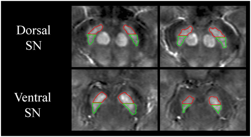
Figure 1. Representative regions of interest drawn on native space QSM images.

Hand-drawn regions of interest (ROIs) of the substantia nigra (SN) are shown overlaid on native space magnetic susceptibility image of a 59-year-old male with Parkinson’s disease. The red ROIs correspond to the anterior SN while the green ROIs correspond to the posterior SN. Measurements were separately made in the dorsal SN (adjacent to the red nucleus) and in the ventral SN (inferior to the red nucleus). The image contrast ranges from −100 to 200 parts per billion.
