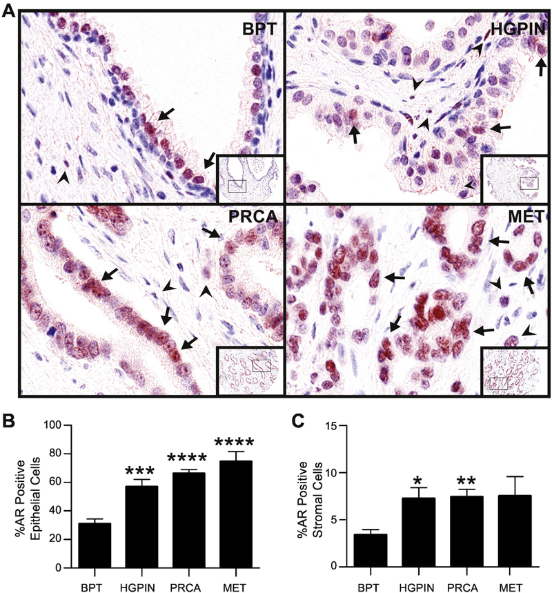
Fig. 1. Androgen receptor expression in prostate cancer progression.
A: Prostate composite image shows androgen receptor (AR, red) in nuclei of luminal epithelial cells (arrows) and stromal cells (arrowheads). AR positivity is seen more in the epithelium than stroma, and overall, expression increased in PRCA progression.
B: The percentage of AR positive cells significantly increased in the epithelium in HGPIN, PRCA and MET compared to BPT.
C: The percentage of AR positive cells in the stroma significantly increased in HGPIN and PRCA compared to BPT.
BPT = benign tissue prostate, HGPIN = high grade prostatic neoplasia, PRCA = prostate cancer, MET = metastasis. IHC 100X (20X inset). *P < 0.05, **P < 0.01, ***P < 0.001, ****P < 0.0001 via post-hoc comparison to BPT.

