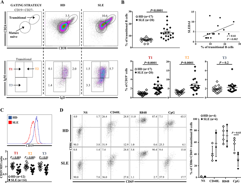
Fig. 1.
(A) Representative dot plots of transitional B cells and their subsets in one HD and one SLE patient. (B) Frequency of transitional B cells (top left panel) and their subsets (bottom panel) (% of CD19+) in HDs compared to SLE patients. Correlation between transitional B cells (% of CD19+) and disease activity assessed by the SLEDAI-2K score (top right panel). (C) MFI of CD19 in transitional B cells subsets from HDs and SLE patients. (D) Frequency of CD86+CD69+ transitional B cells from HDs or SLE patients after no stimulation (NS) or in vitro stimulation with CD40L (CD40 ligand), R848 (TLR7 agonist) or CpG (TLR9 agonist) for 2 days. HDs: healthy donors; MFI: mean of fluorescence intensity; SLE: Systemic lupus erythematosus.

