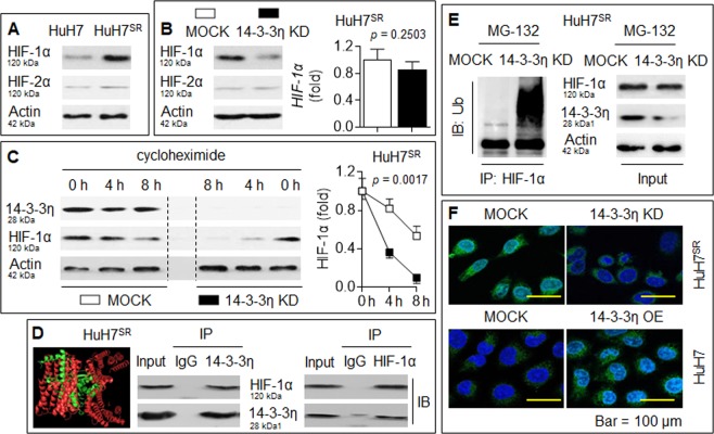
Fig. 2. 14-3-3η stabilized and activated HIF-1α.
a IB analysis of the expressions of HIF-1α and HIF-2α in HuH7 and HuH7SR cells. b HuH7SR cells were transfected by scrambled or 14-3-3η siRNA, IB (left) and qRT-PCT (right) analysis of the expressions of HIF-1α or HIF-2α. c After HuH7SR cells were transfected by scrambled or 14-3-3η siRNA, they were treated by cycloheximide for 0, 4, or 8 h. IB analysis of the expression of HIF-1α. d Computer-docking (left) and IP analysis (right) of the relationship between 14-3-3η and HIF-1α proteins in HuH7SR cells. e After HuH7SR cells were transfected by scrambled or 14-3-3η siRNA, they were treated by MG-132 for 2 h, IP analysis of the ubiquitination of HIF-1α. f HuH7SR cells were transfected by scrambled or 14-3-3η siRNA, while HuH7 cells were transfected by scrambled or 14-3-3η plasmid. Immunostaining analysis of the expression and intracellular distribution of HIF-1α

