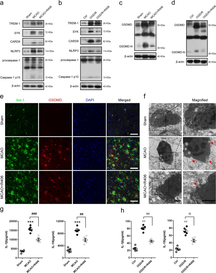
Fig. 9. TREM-1-induced SYK activation triggered microglial pyroptosis post-stroke.
MCAO mice and OGD microglia were treated with SYK inhibitor, R406. a, b Western blotting analysis of TREM-1, SYK, CARD9, NLRP3, and caspase-1 in MCAO mice 3 days after reperfusion and in OGD microglia 24 h after reoxygenation. c, d Immunoblotting analysis for GSDMD in treated mice and cultured microglia. e Double immunostaining of Iba-1 and GSDMD revealed a good co-localization of these two makers. Treatment with LP17 reduced GSDMD positive microglia in ischemic penumbra. Scale bar = 50 µm. f Representative transmission electron microscopy images of microglia in peri-infarct area. Magnified views of microglial cytomembrane are marked with dashed line boxes. Red arrow head: membrance pores. Scale bar = 1 µm. g, h ELISA assays for IL-1β and IL-18 in brain tissues and microglial culture medium. n = 5 in each group. Data are expressed as mean ± SEM. ***p < 0.001 vs. sham group; ##p < 0.01, ###p < 0.001 vs. MCAO group; †††p < 0.001 vs. control group; §§p < 0.01, §§§p < 0.001 vs. OGD/R group

