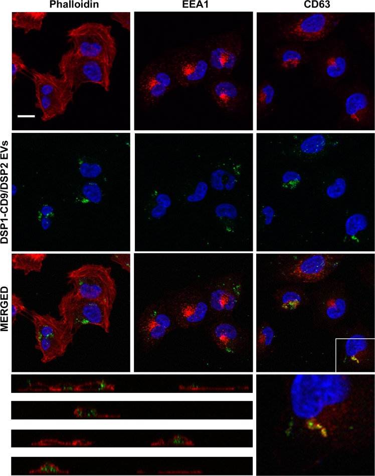
Figure 6.
DSP1-CD9/DSP2 EVs uptake detected by fluorescence confocal microscopy. SUM159 target cells were incubated with DSP1-CD9/DSP2 EVs isolated by ultracentrifugation. Cells were fixed and permebilized after 5 h of incubation with EVs. Samples were stained with Phalloidin-647, αEEA1 antibody or αCD63 (Tea 3/10) mAb. All samples were co-stained with DAPI and analysed in a confocal fluorescence microscope. A maximal projection of the central optical sections is shown. Below, a series of vertical sections of Phalloidin-stained samples are depicted, clearly showing the intracellular localization of uptaken EVs. On the right, a zoomed image in which colocalization of EVs with CD63 labelling is demonstrated on a single confocal plane. Bar = 10 μm.

