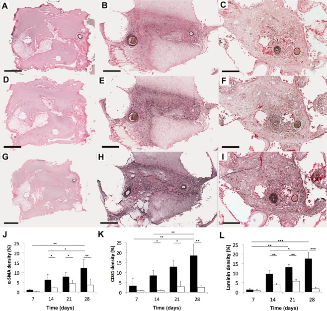
Figure 4.
Sections from AVL chambers for blood vessel markers. (A–C) α-SMA (blood vessel wall), (D–F) CD31 (endothelial cell), (G–I) laminin (basement membrane) were stained at day 7 (left column), day 14 (middle), day 28 (right). Increasing numbers of blood vessels (dark brown structures) appear with increasing time. Scale bar = 500 µm. The areas of staining for (J) α-SMA (K) CD31 (L) laminin were then measured and expressed as a percentage of the total chamber area, mean ± SD in AVL (black) compared to no AVL controls (white) over time. ***P < 0.0001, **P < 0.001, *P < 0.05. Larger magnifications can be found in Supplementary Fig. S5A–F.

