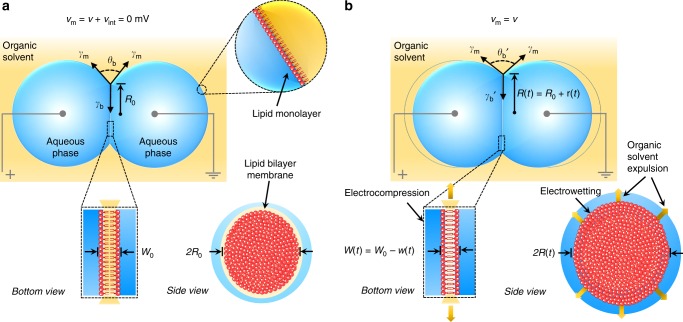
Fig. 1.
Biomimetic membrane assembly and electromechanical behaviours. a A capacitive planar lipid bilayer that mimics the structure of a biological membrane forms spontaneously upon contact between lipid-coated droplets and exclusion of excess oil. The elliptical interface represents an equilibrium in adhesive forces governed by: (1) a balance of monolayer, , and bilayer, tensions prescribed by Young’s equation (Supplementary Note 2, Eq. S.2); and (2) the slight sagging of droplets caused by the water-oil density difference. In the absence of a net membrane voltage, the geometry of the bilayer is described by the zero-volt minor axis radius, R0 (~100–300 µm, determined by analysis of bottom-view, bright-field images (Supplementary Fig. 2), and the hydrophobic thickness, W0 (~2–4 nm). Wire-type (~125 µm diameter) silver/silver chloride (Ag/AgCl) electrodes inserted into the droplets were used to apply a transmembrane voltage and measure the induced ion current. Aqueous droplets (pH 7) contained 500 mM potassium chloride and 10 mM MOPS (3-(N-morpholino)propanesulfonic acid). We define the membrane voltage, vm, as the summation of the applied voltage, v(t), and the intrinsic membrane potential, vint (equal to zero for symmetrical membranes). b A schematic describing the geometrical changes caused by a net membrane voltage, v(t). Changes are manifested by EW-driven creation of new bilayer area between opposing lipid monolayers (at constant thickness) and an independent decrease in hydrophobic thickness due to EC-driven removal of residual oil in the membrane. Since the volumes of both droplets remain constant, the external contact angle, , and bilayer radius, R(t), increase as EW reduces bilayer tension, . Monolayer tension, , is independent of transmembrane voltage (Supplementary Fig. 3)

