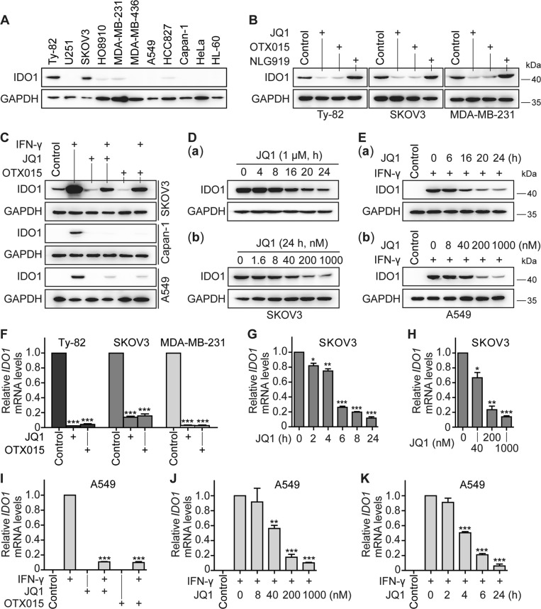
Fig. 2. BET inhibitors reduce the constitutive and IFN-γ-induced expression of IDO1.
The protein levels of IDO1 were detected by western blotting in human tumor cell lines derived from different tissues (a), in Ty-82, SKOV3, and MDA-MB-231 cells treated with JQ1, OTX015, or NLG919 at 1 μM for 24 h (b), in SKOV3, Capan-1, and A549 cells that were treated with IFN-γ (10 ng/ml), JQ1 (1 μM), OTX015 (1 μM), or their respective combinations for 24 h (c), in SKOV3 (d) and A549 (e) cells treated with JQ1 (1 μM) [d(a)] or IFN-γ (10 ng/ml, 24 h) plus JQ1 (1 μM) [e(a)] for the indicated time or with gradient concentrations of JQ1 [d(b)] or JQ1 plus IFN-γ (10 ng/ml) [e(b)] for 24 h. GAPDH was used as the loading control. The relative mRNA levels of IDO1 were determined by RT-qPCR in Ty-82, SKOV3, and MDA-MB-231 cells treated with JQ1 or OTX015 at 1 μM for 24 h (f), in SKOV3 cells treated with JQ1 (1 μM) for the indicated time (g) or with gradient concentrations of JQ1 for 24 h (h), or in A549 cells treated with IFN-γ (10 ng/ml) plus JQ1 or OTX015 (1 μM) for 24 h (i), with IFN-γ (10 ng/ml) plus gradient concentrations of JQ1 for 24 h (j), or with IFN-γ (10 ng/ml, 24 h) plus JQ1 (1 μM) for the indicated time (k). All the data were from three independent experiments, and if applicable, were expressed as mean ± SD (error bar); *p < 0.05; **p < 0.01; ***p < 0.001; + , treated with the indicated drug

