Figure 6.

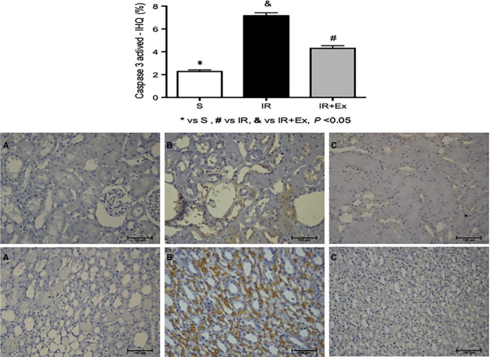
Caspase activation in tubular cells during renal ischemia‐reperfusion injury in sedentary and exercised rats. Male rats sedentary and exercised were subjected to 45 min of renal ischemia followed by 48 h of reperfusion. Renal tissues were fixed for immunofluorescence using an antibody specific for active caspase‐3. (A) Representative images of light marking for caspase‐3 active in renal tubular cells of the sham group. (B) Representative images of less intense marking for caspase‐3 active in renal tubular cells of the EX + IR group. (C) representative images of intense marking for caspase‐3 active in renal tubular cells of the IR group. Values are expressed as means ± SD. * (P < 0.05) versus sham, # (P < 0.05) versus IR, & (P < 0.05) versus EX + IR. [cortex (top) and medulla (bottom); 200×].
