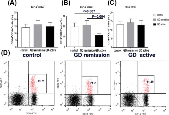
Figure 3.

The percentage of FcγRI (CD64), FcγRII (CD32), and FcγRIII (CD16) on monocytes from Grave' disease (GD) in active and in remission and controls by flow cytometric analysis. A, Bar graphs from three groups depicting cell percentage of CD64‐enriched monocytes (left panels). B, Bar graphs from three groups depicting cell percentage of CD32‐enriched monocytes (middle panels). C, Bar graphs from three groups depicting cell percentage of CD16‐enriched monocytes (right panels). D, Flow cytometry dot plots from three groups depicting CD32 expression by CD14‐enriched monocyte cells. The number in the right quadrants represents the percentage of CD14+CD32+ in each group. Data are presented as the means ± standard error. One‐way ANOVA was used to compare the differences in multiple groups. P < 0.05 was considered significant. GD, Graves' disease
