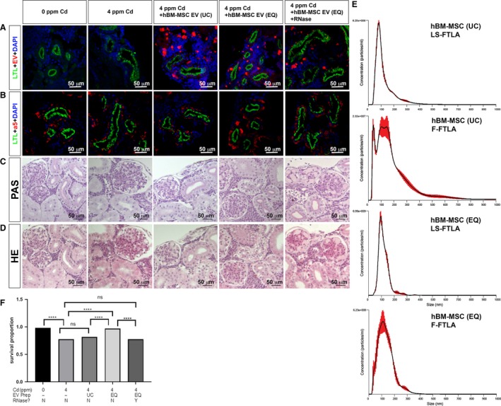
Figure 9.

Human BM‐MSC‐derived EVs repair kidney injury in Cd‐exposed medaka. (A) LTL labeled the PT apical membrane (green). ExoGlow‐membrane red dye labeled hBM‐MSC EVs (red). DAPI was used to stain nuclei (blue). (B) LTL labeled the PT apical membrane (green). LTL (green), DAPI (blue), and a5 labeled the Na+, K+‐ATPase alpha subunit basolateral localization (red). (C) PAS staining of JB4 sections in the kidneys. (D) HE staining of JB4 sections in the kidney. (E) Fluorescence nanoparticle analysis. Finite Track Length Adjustment (FTLA), FTLA measurement for light scattering (LS‐FTLA). Regular size/concentration measurement for light scattering (LS‐SC), FTLA measurement for fluorescent data (F‐FTLA), regular size/concentration measurement for fluorescent data (F‐SC). (F) Survival proportion graph of 7 dai female medaka exposed to Cd and treated with different preparations of BM‐MSC EVs. Significance by Log‐rank test. *****P < 0.0001; ns = not significant. The white scale bar indicates 50 μm (A, B). The black scale bar indicates 50 μm (C, D).
