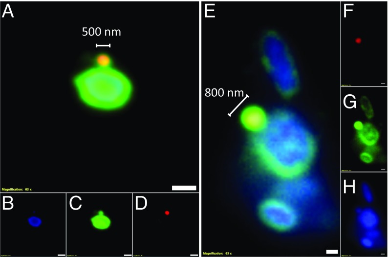
Fig. 5.
FISH of Nha-C FACS cells with Hrr. lacusprofundi ACAM34. Fluorescence micrographs show Nha-C cells in contact with Hrr. lacusprofundi cells. The Nha-C cells fluoresced for both the Nha-C and Hrr. lacusprofundi probes, indicating that Hrr. lacusprofundi rRNA transfers to Nha-C cells. Nha-C cells are labeled with a Cy5-conjugated (red fluorescence) probe. Hrr. lacusprofundi cells are labeled with a Cy3 (yellow fluorescence; recolored to green to improve contrast) probe; all nucleic acid-containing cells are stained with DAPI (blue fluorescence). Composite image of all 3 filters (A and E). Individual filters for Cy3 (B and F), DAPI (C and G), and Cy5 (D and H). (Scale bars: 2 µm.)

