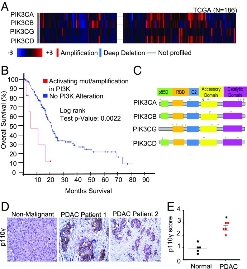
Fig. 2.
PI3K p110γ is up-regulated in patients with PDAC. (A) Copy number alteration (CNA) among four PI3K gene isoforms—p110α (PIK3CA), p110β (PIK3CB), p110γ (PIK3CG), and p110δ (PIK3CD)—in 186 patients with PDAC represented as a heatmap, which was obtained from the provisional TCGA database from cBioPortal (http://www.cbioportal.org). The CNA analysis was performed using putative CNAs from GISTIC2 (Genomic Identification of Significant Targets in Cancer) that assign the following values: −2 = homozygous deletion; −1 = hemizygous deletion; 0 = neutral /no change; 1 = gain; 2 = high level amplification. The heatmap displays log2 copy numbers. (B) Kaplan–Meier survival curve generated by the cBioPortal. Shown are the overall fractions of subjects surviving over time with and without alterations in the four PI3K gene isoforms: PIK3CA, PIK3CB, PIK3CG, and PIK3CD. The alterations include amplifications and activating mutations. A robust difference in survival across groups was observed in the patients with PDAC (P = 0.0022). (C) Identified p110 alterations and their locations in the combined dataset. p85-BD, p85-binding regulatory domain; RBD, RAS-binding domain. (D and E) IHC evaluation of p110γ expression in adjacent nonmalignant and PDAC tumor tissue scored from 0 to 3+ (0, no detectable immunostaining; 1, 10–30% immunostaining; 2, 30–60% immunostaining; 3, >60% immunostaining). n = 5/group; PDAC vs. normal, P = 0.0119.

