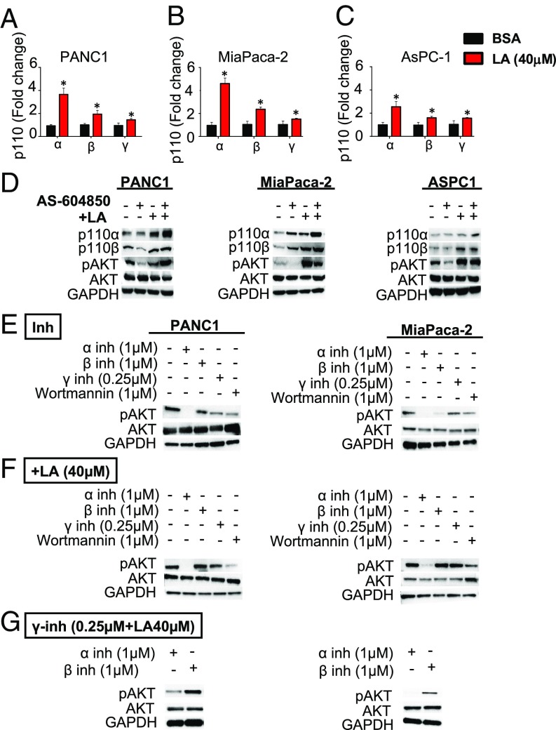
Fig. 6.
Exogenous supplementation of ω-6 fatty acids activates the AKT pathway. (A–C) Panc-1 (A), MiaPaca-2 (B), and AsPC-1 (C) cells were treated with LA (40 μM), and p110α, p110β, and p110γ expression in vitro was evaluated by qPCR. (D) Panc-1, MiaPaca-2, and AsPC-1 cells were treated with LA 40 μM, the p110γ specific inhibitor AS-604850 (0.25 μM), or a combination of the two, and P110α and p110β expression in vitro was evaluated by Western blot analysis, which also shows AKT activation as determined by pAKT levels. (E) pAKT is activated by an alternate p110 isoform after γ-inhibition in the setting of HFD. Panc-1 and MiaPaca-2 were incubated with p110-isoform specific inhibitor p110α (BYL719) or p110β (GSK2636771) for 48 h, and pAKT level was assessed by Western blot analysis. (F) Panc-1 and MiaPaca-2 were then incubated in the presence of LA (40 µM) for 48 h, and pAKT levels were assessed by Western blot analysis. (G) Panc-1 and MiaPaca-2 were then incubated in the presence of AS-604850+LA and either BYL719 or GSK2636771. Increased pAKT was abolished in the presence of BYL719. All assays were performed in triplicate, and the results were averaged. *P < 0.05.

