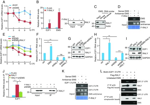
Fig. 4.
EMS cooperates with RALY to promote E2F1 mRNA stability. (A) A549 cells were infected with lentiviruses expressing either control shRNA (shctrl) or EMS shRNA (shEMS). Forty-eight hours later, cells were incubated with actinomycin D (2 μg/mL) for the indicated periods of time. Total RNA was then analyzed by real-time RT-PCR to examine E2F1 mRNA stability. Data shown are mean ± SD (n = 3). (B) A549 cells were infected with lentiviruses expressing pSin-control or pSin-Flag-RALY. Forty-eight hours later, cell lysates were immunoprecipitated with anti-Flag antibody. RNAs in immunoprecipitates (IP) were then analyzed by real-time RT-PCR to examine EMS and E2F1 mRNA levels. Data shown are mean ± SD (n = 3). **P < 0.01. The input and immunoprecipitates were also analyzed by Western blotting. (C) Lysates from A549 cells were incubated with either sense or antisense biotin-labeled DNA oligomers corresponding to EMS, followed by the pull-down experiments using streptavidin-coated beads. The pull-down complexes were analyzed by Western blotting. (D) In vitro-synthesized EMS or its antisense RNA was incubated with purified recombinant Flag-RALY bound with M2 beads. The bead-bound RNAs were then eluted as templates for RT-PCR analysis. (E) A549 cells were infected with lentiviruses expressing control, EMS shRNA, or both EMS shRNA and RALY. Forty-eight hours later, cells were incubated with actinomycin D (2 μg/mL) for the indicated periods of time. Total RNA was then analyzed by real-time RT-PCR. Data shown are mean ± SD (n = 3). (F) A549 cells were infected with lentiviruses expressing control (ctrl), EMS, RALY shRNA (shRALY), or both EMS and RALY shRNA. Forty-eight hours later, total RNA was analyzed by real-time RT-PCR. Data shown are mean ± SD (n = 3). *P < 0.05; **P < 0.01, ns., no significance. (G) A549 cells were infected with lentiviruses expressing ctrl, EMS, RALY shRNA, or both EMS and RALY shRNA. Forty-eight hours later, cell lysates were analyzed by Western blotting. (H) A549 cells were infected with lentiviruses expressing control, EMS shRNA, RALY, or both EMS shRNA and RALY. Forty-eight hours later, total RNA was analyzed by real-time RT-PCR. Data shown are mean ± SD (n = 3). *P < 0.05; **P < 0.01; ***P < 0.001. (I) A549 cells were infected with lentiviruses expressing control, EMS shRNA, RALY, or both EMS shRNA and RALY. Forty-eight hours later, cell lysates were analyzed by Western blotting. (J) A549 cells transduced with lentiviruses expressing either control shRNA or EMS shRNA were transfected with or without Flag-RALY as indicated. Twenty-four hours after transfection, cell lysates were immunoprecipitated with anti-Flag antibody. RNAs present in immunoprecipitates (IP) were then analyzed by real-time RT-PCR. Data shown are mean ± SD (n = 3). *P < 0.05. The input and immunoprecipitates were also analyzed by Western blotting. (K) Purified recombinant Flag-RALY bound with M2 beads was incubated with in vitro-synthesized E2F1 3′-UTR, EMS, and its antisense RNA in the indicated combination. The bead-bound RNAs were then eluted as templates for RT-PCR analysis. (L) In vitro-synthesized biotin-labeled E2F1 3′-UTR and unlabeled EMS plus Flag-RALY were incubated at room temperature for 30 min. The mixtures were first immunoprecipitated with Flag-M2 beads, followed by the elution step with 3 × FLAG peptides. Half of the eluent was subjected to Western blot and RT-PCR analysis. The rest of the eluent was further immunoprecipitated with streptavidin beads. After extensive washing, the immunoprecipitates were analyzed by Western blotting and RT-PCR.

