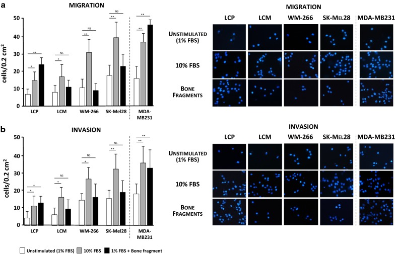
Fig. 1.
Migration and invasion assays of melanoma cell lines. a The migration of melanoma cells was enhanced by 10% FBS as compared to unstimulated cells (average: 25.3 ± 5.5 vs 11.4 ± 2.8 cells/0.2 cm2). The stimulation with bone fragment significantly increased the migration of LCP with respect to unstimulated cells, while produced modest effects on LCM, WM-266 and SK-Mel28. MDA-MB231 breast cancer cells were the positive control. b The invasion assay also confirmed the general invasive attitude under the FBS stimulation (WM-266: 26.3 ± 7.8 cells/0.2 cm2; SK-Mel28: 38.9 ± 9.7 cells/0.2 cm2), although only LCP showed a significant increase of invasiveness (13.1 ± 3.4 vs 4.4 ± 2.9 cells/0.2 cm2) when stimulated with bone fragment as for MDA-MB231 cells (33.6 ± 10 vs 18.7 ± 4.8 cells/0.2 cm2). Bars are mean ± SEM. NS: not significant; *p < 0.05; **p < 0.01. Images on the right side are representative of fluorescence microscopy at ×40 magnification of cells trapped within the trans-well membranes as effect of different stimulations

