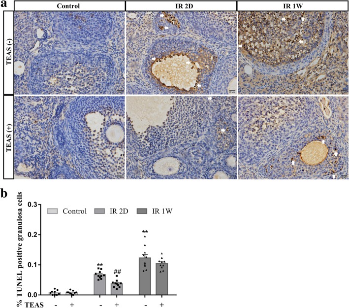
Fig. 6.
In situ end labeling of DNA fragmentation on ovary sections. a Representative image of TUNEL immunostaining in granulosa cells of follicles. b Graphs show the mean ± SEM percentage of follicles with TUNEL-positive granulosa cells. * P < 0.05, **P < 0.01, vs. control- group; # P < 0.05, ## P < 0.01, vs. IR 2D- group. n = 10 mice/group. Arrow: TUNEL-positive granulosa cells, scale bars: 20 μm

