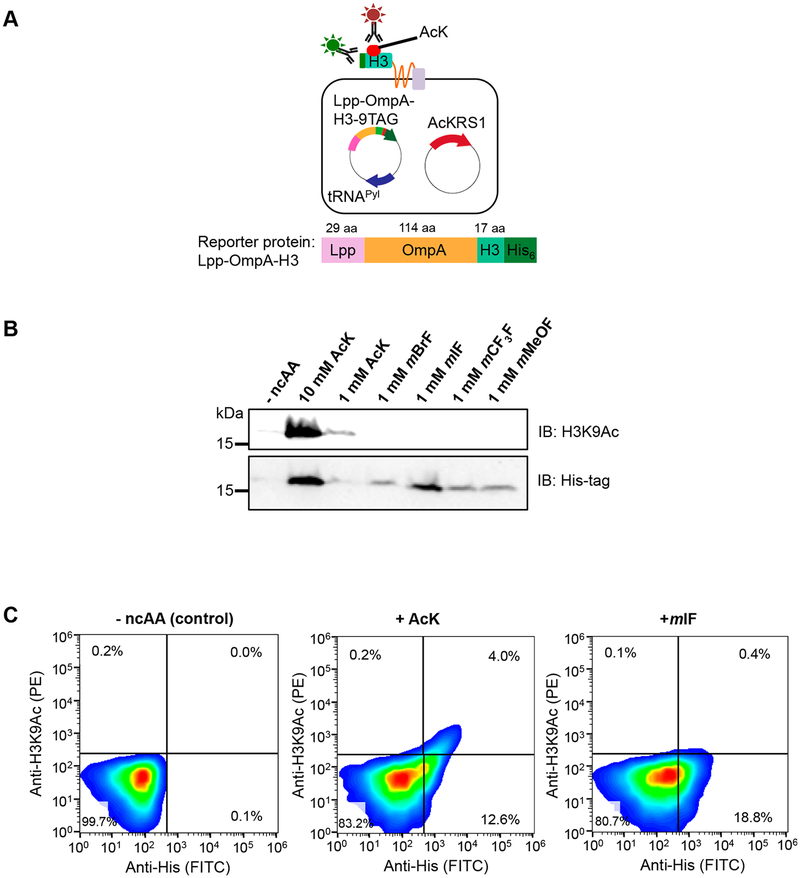
Figure 3.
Specific labeling of AcK-containing Lpp-OmpA-H3 reporter protein allowing selection of engineered tRNA synthetase variants with improved selectivity toward AcK. (A) Presentation of His6-tagged histone 3 (H3) peptide on the surface of E. coli cells by the Lpp-OmpA display scaffold (top). Full-length Lpp-OmpA-H3 can be detected by anti-His6 antibody, and AcK incorporation can be detected by anti-acetyl-lysine antibody in live E. coli cells. Schematic representation of Lpp-OmpA-H3 fusion gene structure (bottom). (B) Anti-acetyl lysine H3K9 antibody specifically detects AcK-incorporated Lpp-OmpA-H3. Western blotting with anti-acetyl-lysine H3K9 (top) for AcK incorporation and anti-His6 for full-length Lpp-OmpA-H3 (bottom). Cells expressing AcKRS1/tRNACUAPyl and Lpp-OmpA-H3 (9TAG) were grown in LB media containing AcK (1 mM or 10 mM), mBrF (1 mM), mIF (1 mM), mCF3F (1 mM), or mMeOF (1 mM). (C) Flow cytometry reveals specific labeling of cells expressing AcK-incorporated Lpp-OmpA-H3. Cells expressing AcKRS1/tRNACUAPyl and Lpp-OmpA-H3 (9TAG) were grown in LB media containing AcK (10 mM) or mIF (1 mM) and labeled with anti-acetyl-lysine H3K9 (indicated by PE stain) and anti-His6 (indicated by FITC stain) antibodies. Representative fluorescence density plots showing live E. coli cells expressing Lpp-OmpA-H3 with no ncAA (left), AcK (middle), or mIF (right) incorporation. At least 2 × 10⁴ cells were analyzed and presented on each FACS plot.

