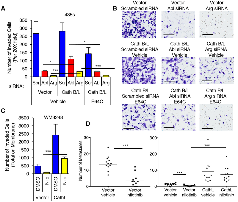
Fig. 8. Abl/Arg drive invasion and metastasis by regulating cathepsin secretion.
(A,B) 435s cells, stably expressing vectors or cathepsin B and L, were transfected with siRNAs (#1), serum-starved, and utilized in a matrigel invasion assay (1% FBS chemoattractant) in the absence or presence of the cysteine cathepsin inhibitor, E64C (50μM; 24h). An aliquot of cells were lysed and blotted (Supplementary Fig. S14A). (B) Representative fields from A. Size bars=100μm. (C) WM3248 cells stably expressing vector or cathepsin L were serum-starved, treated with nilotinib (2μM; 16h), and utilized in a matrigel invasion assay (IGF-1: 10nM; 36h; bottom). An aliquot of cells was lysed and blotted (Supplementary Fig. S14B), and representative fields are shown in Supplementary Fig. S14C. (A,C) Graphs are Mean±SEM, n=3. ***p<0.001; **p≤0.01; *p<0.05 using two sample t-tests and Holm’s adjustment for multiple comparisons. (D) Quantitation of GFP-positive lung nodules from mice injected i.v. with WM3248 cells stably expressing vector or cathepsin L, treated with vehicle or nilotinib for 33 days. Vector-vehicle=n=13 mice; vector-nilotinib, n=11 mice; CathL-vehicle, n=12 mice; CathL-nilotinib, n=11 mice. ***p<0.001, using Wilcoxon rank sum test and Holm’s adjustment for multiple comparisons. (Left) Vector groups. (Right) All treatment groups.

