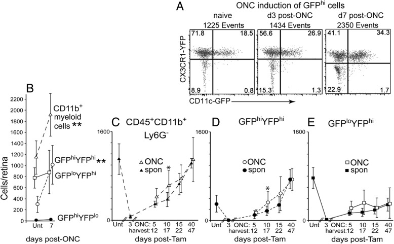
Fig. 4.
The injury response of the repopulating retina after tamoxifen-induced DTA depletion was diminished compared to controls. CD11cDTR/GFP:CX3CR1YFP-creER:ROSADTA mice were given tamoxifen (2.5 mg on three alternating days) or left untreated (Unt, no tamoxifen) and then given an ONC and harvested at the indicated day post-tamoxifen with retinas analyzed by flow cytometry. a Appearance of GFPhi cells in substantial numbers at 7 days post-ONC in untreated (no tamoxifen) mice. b Quantification of the retinal myeloid cell response to an ONC in untreated mice. Open circles indicate GFPhiYFPhi cells and open squares indicate GFPloYFPhi cells. c–e The injury response was strongly attenuated in tamoxifen-induced DTA-ablated retinas, even if performed after nominal repopulation. The analyzed cell populations include total CD45+CD11b+Ly6G− cells (c), GFPhiYFPhi cells (d), and GFPloYFPhi cells (e). Cell numbers are given as mean ± SD with 4–8 samples at each time point. Ipsi-ONC (tamoxifen plus ONC, open symbols) and spontaneously (spon) recovering retinas (tamoxifen only, closed symbols) indicated. *P < 0.05; **P < 0.01 for ONC versus non-ONC retinas

