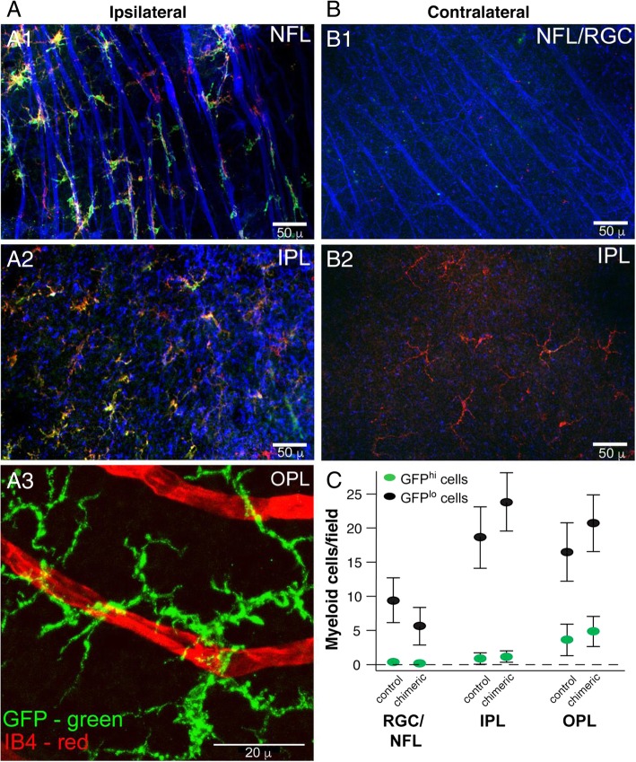
Fig. 9.
Injury response and long-term repopulation of donor-derived myeloid cells recruited into the retina mimics that of endogenous microglia. CD11cDTR/GFP bone marrow was grafted into irradiated (2 × 6 Gy, unshielded) B6 mice. a Analysis of the ipsilateral (ONC injured) retina from a chimeric mouse given an ONC 6 weeks post-bone marrow transfer and analyzed 7 days post-ONC. A1 Staining of retinal ganglion cells (RGC) and their axons in the nerve fiber layer (NFL) and underlying inner. A2 and outer A3 plexiform layers (IPL, OPL). All three layers analyzed were from the same microscopic field of the retina. b Same mouse as a. Analysis of the NFL/RGC and underlying IPL in the contralateral (uninjured) retina. For A1, A2, B1, and B2, green—GFP, blue—β3 tubulin, red—CD11b. For A3, green—GFP, red—isolectin B4 (IB4). c Analysis of myeloid cells in retinal layers in age-matched normal (control) CD11cDTR/GFP mice and chimeric mice 180 days post-bone marrow transfer without ONC. Results are given as the mean number of cells per field ± SD, n = 4 (1 retina from four individual mice for each group), P > 0.05 control versus chimeric mice for each retinal cell layer

