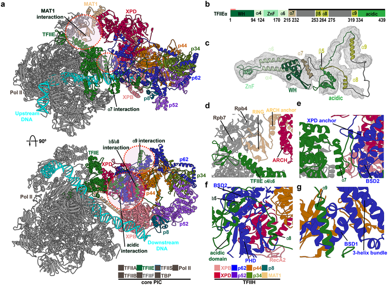
Figure 3. TFIIE, MAT1 and p62 are critical for the integrity of the core-PIC–TFIIH interface.
a, Human PIC structure in cartoon representation with colored TFIIH subunits. Circles demark zoomed regions in d.-g. b, Domain schematic of TFIIEα. c, TFIIEα cartoon. d, MAT1 - core PIC interaction. The MAT1 RING-finger docks into a groove between the Pol II stalk subunit Rpb7 and TFIIE α4-α7 helices. The RING-finger connects to the ARCH anchor which binds the XPD ARCH domain. e, TFIIEα helix α7 is wedged between TFIIE winged helix (eWH) domain and p62. f, TFIIE β5–α7 and acidic domain interacts with p62 PHD and BSD2. g, TFIIE helix α9 binds p62 BSD1 domain adjacent to the p62 3-helix bundle.

