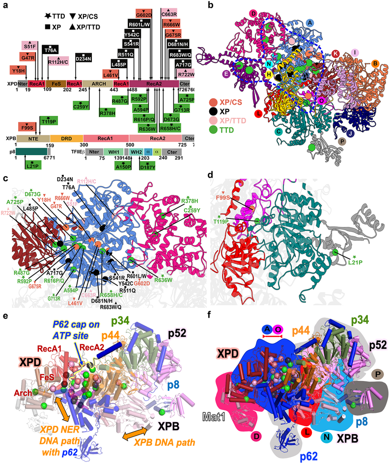
Figure 6. Human disease mutations mapped onto TFIIH and TFIIE show distinct patterns within protein-protein and community interfaces.
a, TTD, XP/TTD, XP and XP/CS point mutations mapped onto XPD, XPB, and TFIIE protein schematic do not co-localize by disease on primary sequence. b, Map of human disease mutations (spheres) onto TFIIH structure as cartoon colored by community show biased localization (blue outline). c, Zoom view of mutations on XPD. d, Zoom view of XPB and p8 mutations. e, Mutations and function mapped onto TFIIH cartoon colored by subunit. Regions of p62 and p44 are removed for clarity. f, Overlay of disease mutations, protein chain (cartoon view), and communities (background color). View matches e.

