
Fig. 2.
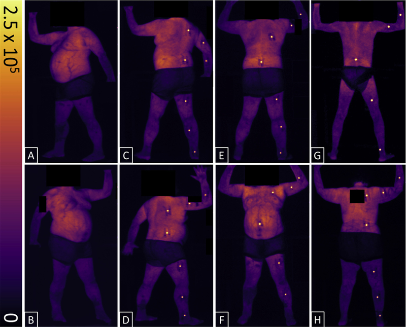
Cumulative, background-subtracted, Cherenkov, and scintillation-intensity maps for patients undergoing total skin electron therapy. All 6 total skin electron therapy positions are shown for 1 patient: left anterior oblique (A), right anterior oblique (B), left posterior oblique (C), right posterior oblique (D), posterior-anterior (E), and anterior-posterior (F). Sample images from the 2 other patients in the posterior—anterior position are shown in G and H. Color-intensity scale is in digital units. Dark areas around the midsection of patients are caused by cloth shorts; all identifying features have been anonymized.