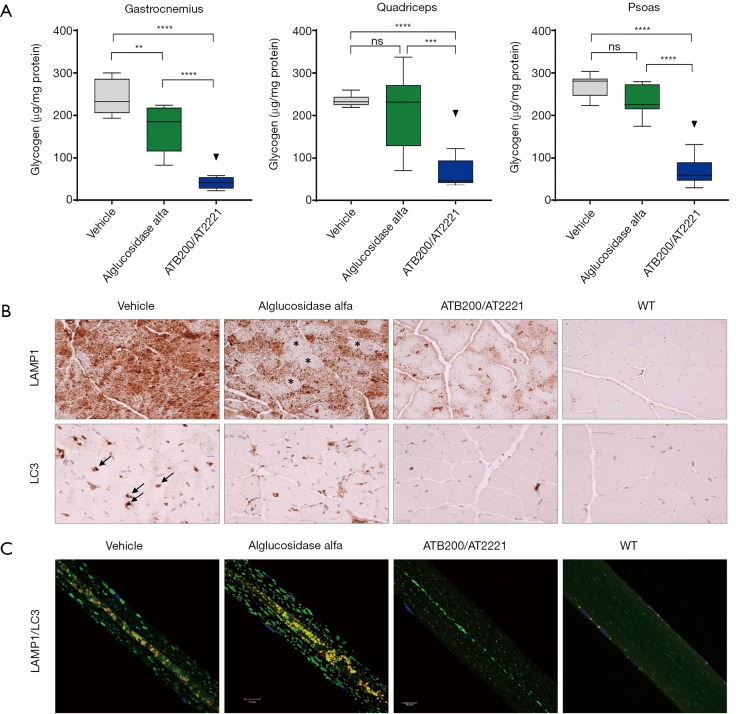
Figure 4.
Approximately 16-week-old male Gaa KO mice received four biweekly intravenous (IV) administrations of vehicle, 20 mg/kg alglucosidase alfa, or 20 mg/kg ATB200/AT2221 (10 mg/kg AT2221 was administered orally 30 minutes prior to ATB200 IV injection). Tissues were collected 14 days after the last administration. (A) Glycogen levels in different skeletal muscles n=6–8 animals per group; ns: not significant (P>0.05); **, 0.001<P<0.01; ***, 0.0001<P<0.001; ****, P<0.0001; Tukey’s multiple comparison under one-way ANOVA. (B) Lamp1 (lysosome-associated membrane protein 1, lysosomal marker, upper panel) and LC3 (microtubule-associated light chain protein, autophagosomal marker, lower panel)-stained sections of skeletal muscle (quadriceps). N=4–5 animals per group (n=2 for the WT), the scale bars: 50 µm. (C) Immunostaining of single fibers from the white part of gastrocnemius with markers for lysosomes (Lamp1; green), autophagosomes (LC3; red), and nuclei (Hoechst dye; blue); the multicolored areas in the core of muscle fibers represent autophagic buildup. n=141 fibers from 4 alglucosidase alfa-treated Gaa-KO mice; n=127 fibers from 4 ATB200/AT2221-treated Gaa-KO mice, the scale bars: 20 µm.

