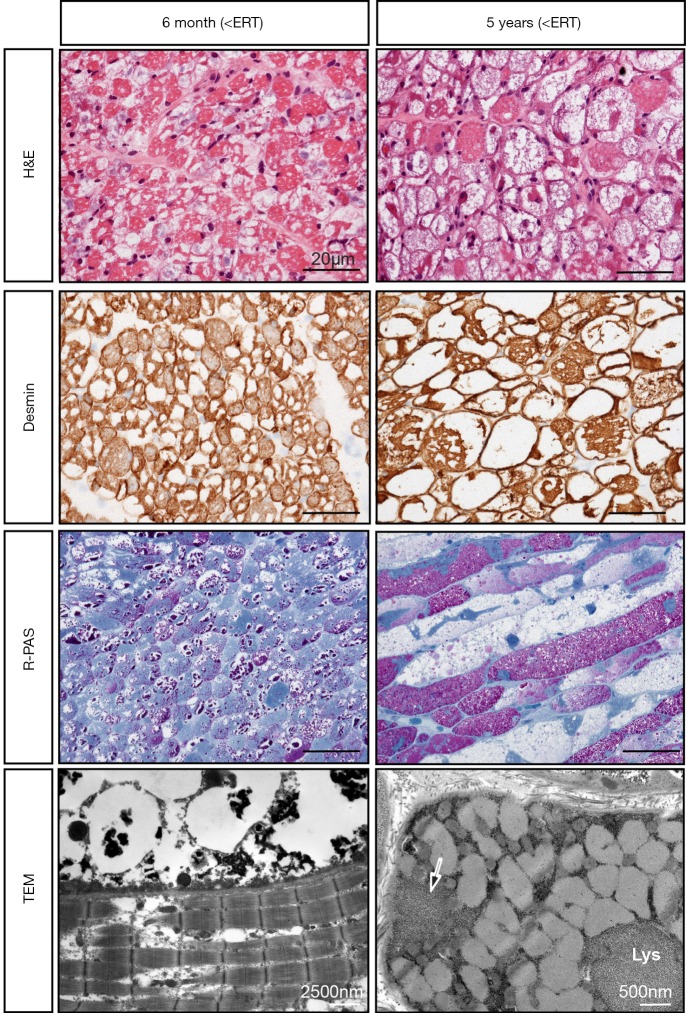
Figure 2.
Muscle biopsy findings (right lateral vastus muscle) in a CRIM-positive patient with IOPD achieving free walking at age 19 months and still doing so at age 8 years. Biopsies were taken at age 6 months before start of ERT (left panel) and at age 5 years (right panel). H&E staining shows a progressive vacuolar myopathy, while Desmin staining depicts absent desmin in many fibers, reflecting increasing myofibrillar damage. Resin-PAS semithin (R-PAS) sections show distinct glycogen accumulation (stained red) at age 6 months and persistence of such fibers in conjunction with fibers full of empty vacuoles mirroring abnormal autophagy at age 5 years. Notice that fibers being in different stages of disease pathology are located side by side. Electron microscopy (TEM) depicts a fiber with normal myofibrils (lower part of the image) and a completely destroyed one (upper part of the image) at age 6 months, and a fiber with normal myofibrills, but marked lysosomal (Lys) and extralysosomal (arrow) glycogen deposits. CRIM, cross-reactive immunological material; IOPD, infantile-onset Pompe disease; ERT, enzyme replacement therapy. Length of the scale bar in is 20 µm in all light microscopic images.

