Figure 3.
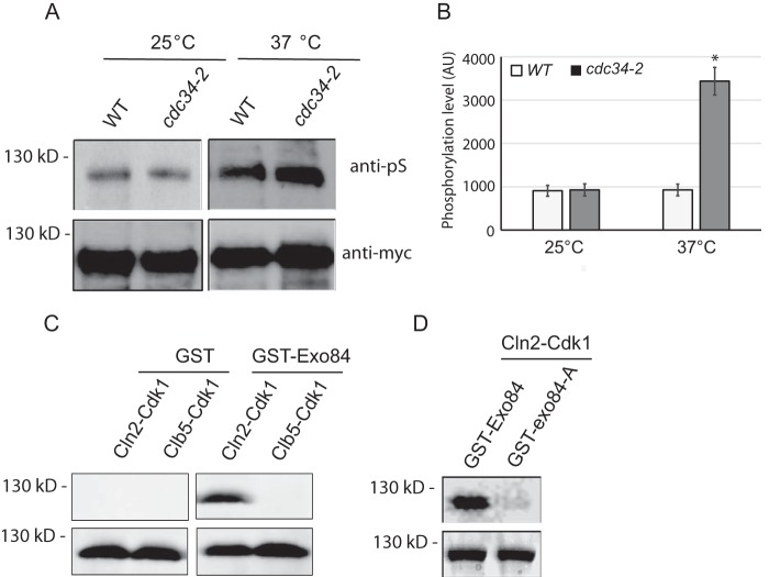
Exo84 is phosphorylated by Cdk1 at late G1 phase. A, Exo84-myc was immunoprecipitated from the WT and cdc34-2 mutant cells at the permissive or restrictive temperature and then probed for Cdk1 phosphorylation by immunoblotting. B, quantification of Exo84 phosphorylation level in A. Error bars represent S.D. (n = 3). *, p < 0.01. C, Exo84 is phosphorylated by Cdk1 in vitro. GST and GST-Exo84 were purified from E. coli and incubated with Cln2–Cdk1 or Clb5–Cdk1 in the presence of [γ-32P]ATP. Exo84 phosphorylation was detected by autoradiography (top). Corresponding Coomassie Blue–stained gels are shown on the bottom. D, in vitro Cln2–Cdk1 kinase assays with recombinant Exo84-A, which lacks the five Cdk1 phosphorylation sites. The phosphorylation of Exo84-A by Cln2–Cdk1 is barely detectable. AU, arbitrary unit.

