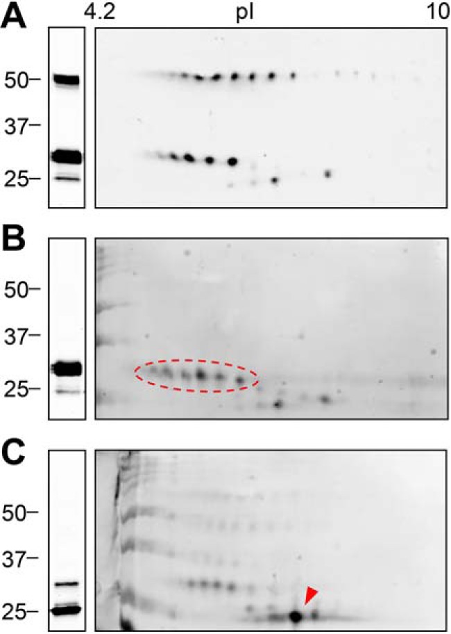
Figure 6.

Analysis of LOX proteolysis by two-dimensional electrophoresis coupled to immunoblotting. LOX supernatants kept under basal conditions (A) or incubated with BMP1 (B) or ADAMTS2 (C) were fractioned by 1D (left) or 2D (right) electrophoresis and analyzed by immunoblotting using a specific C-terminal LOX antibody. LOX precursor was visualized as a train of spots with the isoelectric point (pI) more acidic than predicted from the amino acid sequence (7.99). BMP1-cleaved mature form gave a similar pattern (indicated by a red dashed oval), whereas the cleavage by ADAMTS2 resulted in a single prominent spot at a more basic pI (red arrowhead). The blots shown correspond to representative experiments performed twice with two independent preparations.
