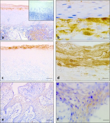
Fig. 2.
Immunohistochemical staining for RNase 7 in oral lichen planus and radicular cyst. Intense immunostaining was observed in the surface layers and granular layers of oral lichen planus (a) and (b). Negative control showed negative staining (inset of a). Strong immunoreactions were observed in the surface layers, pyknotic nuclei and positive dots for RNase 7 (c) and (d). Faint staining for RNase 7 in the epithelial lining of the radicular cyst (e) and (f). Bars = 100 μm (a, c, e); 100 μm insets of (a); 10 μm (b, d & f).

