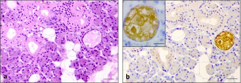
Fig. 4.
H/E staining and immunohistochemical staining for RNase 7 in sebaceous glands often observed in non-inflamed salivary glands. H/E staining showed a pale staining of cytoplasm with shrunken or absence of nuclei in the mature sebaceous cell at the center of sebaceous gland. Immature sebaceous cells at the periphery of sebaceous gland showed rounded nuclei (a). Intense immunoreaction for RNase 7 in the cytoplasm of the sebaceous glands (b) & inset of (b). Bars = 100 μm (a & b); 10 μm insets of (b).

