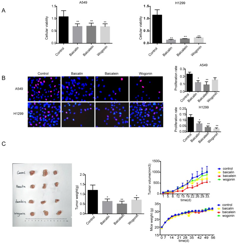
Figure 1.
Baicalin, baicalein and wogonin inhibited the growth of NSCLC in vitro and in vivo. (A) A549 and H1299 were exposed to baicalin, baicalein or wogonin in concentration of 200 μM, 10 μM or 40 μM respectively for 24 h, and cell viability was determined by MTT assay. (B) A549 and H1299 were exposed to baicalin, baicalein or wogonin in concentration of 200 μM, 10 μM or 40 μM respectively for 24 h, cells were incubated with 10 μM EdU for 2 h and EdU assay was performed. Five images of each well and three duplicated wells were taken by fluorescence microscope and EdU positive cells were counted. (C) Balb/c nude mice bearing palpable A549 xenografted tumors were intragastrically administered with control (CMC-Na), baicalin (80 mg/kg), baicalein (40 mg/kg) or wogonin (80 mg/kg) daily. Representative tumors and tumor weights dissected on day 28 after treatment were shown (left). The tumor volumes and mice weight measured twice a week versus time was plotted (right) (n=8). Each bar represented the mean ± SEM. Significant differences were shown (*p < 0.05 and **p < 0.01, compared with the control group).

