
Figure 2:
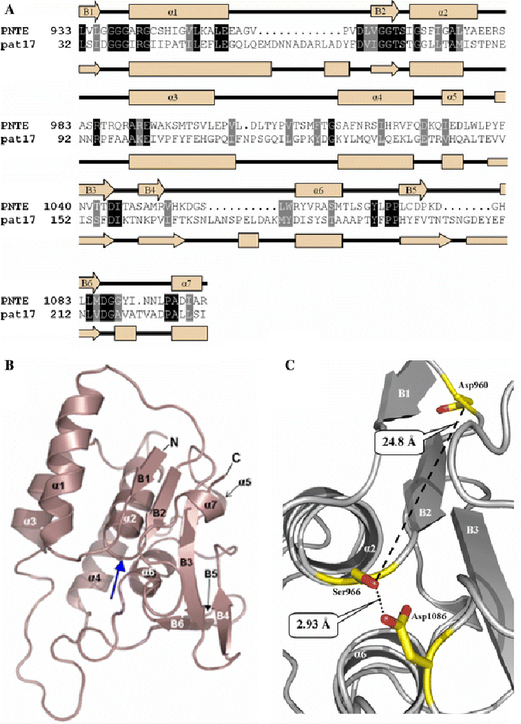
A.) Sequence homologies and identities between PNTE and pat17 sequence alignment of PNTE and pat17. Identical residues are boxed in black, while homologous residues are in grey. Secondary structures of PNTE and pat17 are shown along with their respective primary structures as cream arrows (representing β-strands) and orange rectangles (representing α helices). α-helices and β-strands in the PNTE sequence are numbered α1-α7 and B1-B6, respectively. Shading for homologous and identical residues in the aligned sequences was performed using BoxShade 3.21 (http://www.ch.embnet.org/software/BOX_form.html). B.) A ribbon diagram of the putative tertiary structure for the NTE patatin domain. Blue arrow denotes the location of nucleophilic elbow. C.) Organization of the predicted active site of NTE. Ser966, Asp960, and Asp1086 are depicted as balls-n-sticks. Distances between Ser966 Oγ and Asp960 Oδ2 and Ser966 Oγ and Asp 1086 Oδ2 are shown as dotted lines. Images b. and c. were produced in PyMol.