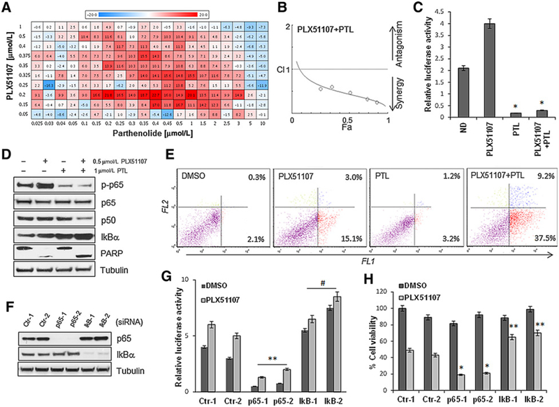
Figure 1.
Parthenolide has synergistic activity in combination with PLX51107 in a high-throughput drug screening. A, Uveal melanoma cells (92.1) were treated in secondary screenings with increasing concentrations of PLX51107 (0.05–1 μmmol/L) and parthenolide (0.025–10 μmmol/L) for 72 hours. The Bliss values for positive interactions (additive/synergy) are indicated in red and negative interactions in blue. B, Chou–Talalay plot (x-axis, Fa, or fractional activity, reflects the fraction of cells affected by the drug treatment relative to vehicle controls; y-axis, combination index, with <1, >1, and = 1 indicating synergistic, antagonistic, and additive effects, respectively). Each point represents a different combination of drug concentrations. C, The cells were transfected with a vector containing an NF-κB reporter gene along with a Renilla luciferase vector (1:10 ratio), then treated with 0.5 μmmol/L PLX51107, 1 μmmol/L PTL, and the combination for 24 hours. Luciferase activity was determined by chemiluminescence. Results are normalized to Renilla luciferase activity and represent the mean ± SD.*, P < 0.005.D, Western blot analysis of 92.1 cells treated with 0.5 μmmol/L PLX51107 and 1 μmmol/L PTL alone and in combination for 48 hours, showing inhibition of p65 phosphorylation, decreased expression of p50, induction of IkBα, and PARP cleavage. E, Apoptosis assay of 92.1 cells treated with 0.5 μmmol/L PLX51107 and 1 μmmol/L PTL alone and in combination for 48 hours, measuring cell permeability to fluorescent stains YO-PRO (FL1, for early apoptosis) and propidium iodide (FL2, for late apoptosis). F, Immunoblotting of cells transfected with two control (siCtr-1, −2), two p65 (sip65–1, −2), and two IkBα (IkB-1, −2) siRNA. G, The siRNA-transfected cells were then transfected with the NF-κB reporter gene vector/Renilla and tested for luciferase activity.**, P < 0.01; #, P < 0.05, comparing sip65–1,−2 with control siRNA. H, Cell viability assays of cells depleted of the indicated proteins after treatment with PLX51107 for 72 hours. Bars, mean ± SD.*, P < 0.005; **, P < 0.01.

