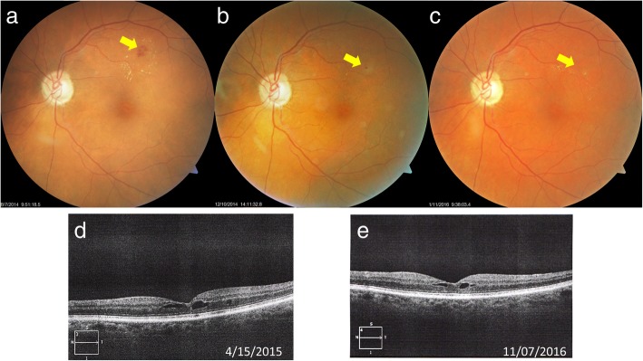
Fig. 3.
Follow-up of third case fundus photos. Some exudate leakage was found at the August 2014 visit (a). After taking Eyefolate™, the large MA (yellow arrow) was reduced and the exudate was smaller at the Dec 2014 visit (b). The MA and exudates (yellow arrow) were resolved by the January 2016 visit (c). In addition, at the November 16 visit (e), the diabetic cystic macular edema was reduced from the 2015 visit (d) and later visit

