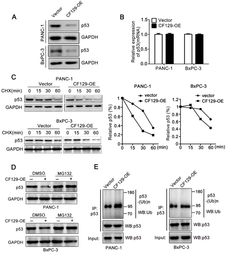
Figure 5.
CF129 induces p53 ubiquitination and degradation. (A) p53 protein levels were assessed in PANC-1/BxPC-3 cells transfected using CF129-OE by Western blot analysis. (B) qRT-PCR assessment of p53 levels in PANC-1/BxPC-3 cells transfected with CF129-OE. (C) After transfected with CF129-OE, CHX (100 ug/mL) was used to treat PANC-1/BxPC-3 cells prior to western blotting, with ImageJ used to quantify p53 band densitometry. (D) After transfected with CF129-OE, PANC-1 and BxPC-3 cells were treated with MG132(20nM) for 3h and used for western blotting. (E) The CF129-OE transfected PANC-1 cells were treated using MG132(20nM) for 3h. Anti-p53 was then used for immunoprecipitation of these cell lysates, followed by western blotting to assess endogenous p53 ubiquitination.

