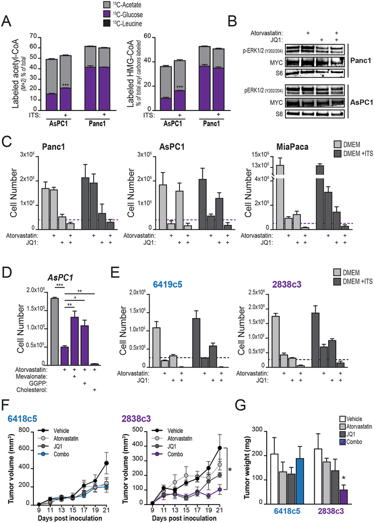
Figure 7: Targeting acetyl-CoA-dependent processes can suppress PDA growth.
A, AsPC1 or Panc1 cells were cultured overnight in DMEM with or without ITS+ (insulin-based supplement, BD Biosciences) and then labeled for 2 hours with indicated substrates (n=3, each condition). Percent labeling of acetyl-CoA and HMG-CoA were determined by LC-MS. Stars denote statistically different labeling from glucose. B, Western blotting shows levels of MYC and ERK1/2 phosphorylation in AsPC1 and Panc1 cells treated with atorvastatin (20 μM) and/or JQ1 (500 nM) for 4 days. C, indicated cell lines were cultured in DMEM with or without ITS+ and treated with either atorvastatin (20 μM), JQ1 (500 nM), or both for 4 days. Graphs show final cell counts. Dashed purple lines denote starting cell number (counted at day 0). Experiments are representative of 2 independent biological repeats. D, AsPC1 cells were treated with atorvastatin (20 μM) and counted after 4 days. Effect of supplementation with mevalonate (100 μM), geranylgeranyl pyrophosphate (GGPP, 100 μM) or cholesterol (12.5 μg/mL) is shown. Cholesterol was tested over a range of concentrations from 5–100 μg/mL and in all cases failed to rescue proliferation in the presence of atorvastatin (only 12.5 μg/mL data is shown). E, KPCY-derived mouse cell lines were cultured in DMEM with or without ITS+ and treated with either atorvastatin (20 μM), JQ1 (500 nM), or both for 4 days. Graphs show final cell counts. Dashed purple lines denote starting cell number (counted at day 0). Experiments are representative of 2 independent biological repeats. F, growth of two KPCY-derived tumor cell clones (6419c5, left; 2838c3, right) implanted subcutaneously into immune-competent C57Bl6/J mice and treated with either atorvastatin (10 mg/Kg), JQ1 (50 mg/Kg), or both, once a day after tumors became palpable (day 9 post-inoculation). G, tumor mass weight (same in F) after excision post-mortem. For panels A-E, bars show mean, +/− SD; for panels F-G, bars show mean +/− SEM (*, p<0.05; **, p<0.01; ***, p<0.001).

