
Figure 1.
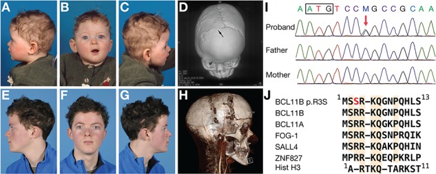
Clinical features. (A–C) Pre-operative facial photographs and 3D-CT scan (D) at the age of 10 months. Right coronal synostosis is indicated by the arrow. (E–G) Post-operative facial photographs at the age of 15 years. (H) 3D-CT and angiogram scan at the age of 12 years just prior to distraction. (I) DNA sequence analysis of proband and parents showing a de novo C>A variant (red arrow) in BCL11B (c.7C>A). The box encloses the start codon. (J) Alignment of amino termini of BCL11B, transcription factors harbouring related sequences and histone H3.