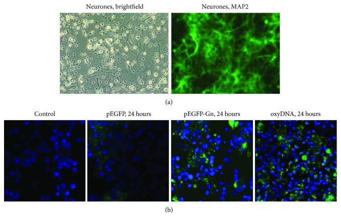
Figure 2.
(a) MAP-2 detection in primary cell culture of Wistar rat cerebellum. MAP-2 (FITC) in neurons. Nuclei are stained with DAPI. ×40. (b) Staining of primary cell culture of Wistar rat cerebellum cells with the various types of the labeled DNA fragments. From left to right: control, pEGFP, pEGFP-Gn, and DNAoxyGreen. Nuclei are stained with DAPI. ×40.

