Figure 6.
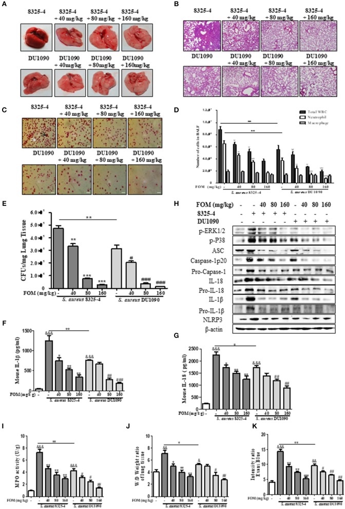
FOM protects mouse S. aureus pneumonia in vivo. (A,B) Gross pathological changes (A) and histopathology (B) of S. aureus-infected lung tissue with or without FOM treatment. Tissues were stained with HE (10×). (C) Wright's-Giemsa stained smear of peritoneal fluid from infected mice with FOM treatment. Neutrophil (white arrow) and Mϕ (black arrow) are presented (20×). (D) The total of WBC, neutrophil and Mϕ in the bronchoalveolar lavage (BAL) fluid of infected mice with FOM treatment were counted stained with Wright's-Giemsa. (E) Bacterial burden in the lungs of infected mice with or without FOM treatment ψ p < 0.05, ψψ p < 0.01, ψψψ p < 0.001 compared with 8325-4 treated group. (F,G) The level of IL-1β and IL-18 in the bronchoalveolar lavage (BAL) fluid of infected mice with FOM treatment was detected by Elisa. (H) The levels of p-ERK, p-P38 and NLRP3 inflammasome protein in S. aureus 8325-4/DU1090-infected lungs with FOM treatment. (I) Effects of FOM on MPO activity of S. aureus -induced lung inflammation in mice. (J) Lung water content was calculated as the ratio of wet weight to dry weight, (K) vascular leakage in lung tissue was measured via injecting Evans blue dye. & p < 0.05, && p < 0.01, &&& p < 0.001 compared with Control group, * p < 0.05, ** p < 0.01, *** p < 0.001 compared with 8325-4 treated group, # p < 0.05, ## p < 0.01, ### p < 0.001 compared with DU1090 treated group. Data are means ± standard errors derived from three experiments.

