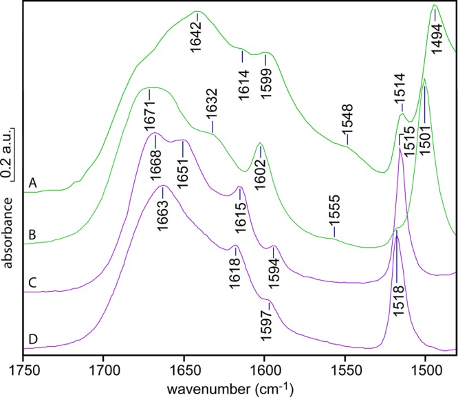
Figure 6.

IR spectra of (p-phenolyl)dodecylguanidine, as the “free base” (green; A, B) or as the HBr salt (purple; C, D), with the former two showing clear evidence of H-bonding and/or proton transfer from the phenol. Each sample was recrystallized from methanol, dried, and then redissolved to a concentration of ∼100 mM in DMSO (A, C) or methanol (B, D). The absorbance scale on the y axis is approximate; the measured spectra had somewhat different concentrations and path lengths and were rescaled for optimum visual comparison.
