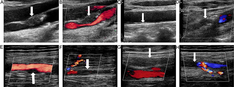
Figure 1. Ultrasonography of the carotid and lower limb arteries of a 74-year-old male patient with diabetic foot, peripheral arterial disease, and plaques in both internal carotid arteries. A, High-resolution gray-scale sonography revealed a plaque (arrow) in the front wall of the right internal carotid artery, and C, an increase in the intima-media thickness of the left common carotid artery (arrow; the measured value was 1.07 cm). B, Color Doppler ultrasound showed severe stenosis of the lumen of the right internal carotid artery (arrow; the diameter was reduced by 70–99%), D, indicated that the left internal carotid artery was occluded (arrow), E, moderate stenosis of the right superficial femoral artery (arrow; the diameter was reduced by 50–99%), F, segmental occlusion in the anterior tibial artery (arrow) along with a collateral artery, G, occlusion of the left superficial femoral artery (arrow), and H, occlusion of the left superficial femoral artery (arrow) accompanied by a collateral artery. The overall ultrasonic score was 10 points for the right lower limb and 9 points for the left lower limb.

