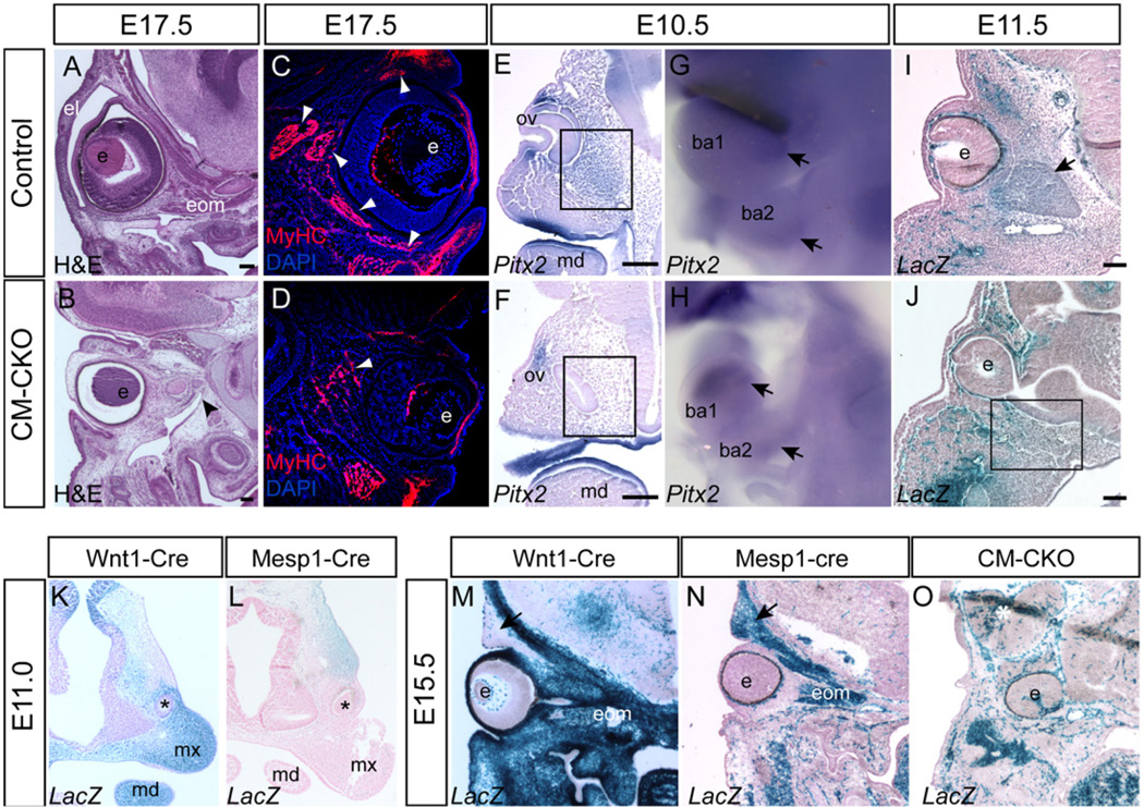
Fig. 3.
Impact of loss of Twist1 in the cranial mesoderm on the development of peri-ocular structures. (A, B) The extra-ocular muscles (eom) are severely underdeveloped in the E17.5 CM–CKO embryo (B, black arrowhead), compared to the age-matched control (A). (C, D) Sparse and loosely organized myosin-heavy chain-positive fibers in the peri-ocular tissue of the E15.5 CM–CKO embryo (D), contrasting with the fiber bundles in the extraocular muscles of the control embryo (C, white arrowheads); the upper eyelids of the CM–CKO embryo (D) are rudimentary and lack muscle fibers. (E, F) In situ hybridization for Pitx2 expression at E10.5 showing reduced presence of Pitx2-expressing cells in the CM–CKO embryo (F) in the area ventral to the eye invagination compared to control embryo (E, boxed area), indicating that the extraocular muscle progenitor population is reduced. (G,H) Pitx2 expression is maintained (black arrow) in the first and second branchial arches of the CM–CKO (I,J) Staining of Mesp1-Cre; Rosa26R cells at E11.5. show a reduced population of Mesp1-Cre; Rosa26R positive mesoderm cells in the tissues surrounding the optic cup and the adjacent orbital area in the CM–CKO (J) compared to the control (I, black arrow, muscle anlagen). (K,L) CM and CNC contribution to the eyelids at E11.0. The Wnt1-Cre; Rosa26R positive CNC cells contributes to the tissues ventral to the eye primordium (asterisk, K) whereas the Mesp1-Cre; Rosa26R positive mesoderm cells to the tissues dorsal to the eye primordium (L). (M–O) Lack of eyelid contribution from the Twist1 deficient CM in the CM–CKO. The contribution of the Wnt1-Cre; Rosa26R positive neural crest cells to the lower eyelid (M), and tissues around the eye and in the neurocranium, compared to that of the Mesp1-Cre; Rosa26R positive mesoderm cells, which contribute to the upper eyelid (black arrow) and extraocular and facial muscles. At E13.5 (O) eyelids and extraocular muscles are absent in the CM–CKO embryo, and there is paucity of Mesp1-Cre; Rosa26R positive mesoderm cells around the eye that is displaced deep in the head (O). ba1, 1st branchial arch; ba2, 2nd branchial arch e, eye; el, eyelid; mx, maxilla; md, mandible; ov, optic vesicle. Scale bar=100 µm.

