Fig. 6.
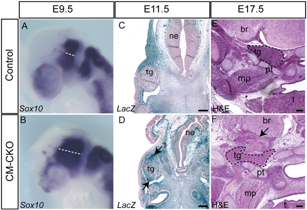
Impact of the loss of Twist1 in the cranial mesoderm on cranial neural crest cells. (A, B) Sox10-expressing neural crest cells remain segregated into pre- and post-otic streams but are more widely spread in the CM–CKO embryo at E9.5 (B) compared to the control (A, dashed line). (C, D) Staining for Rosa26R activity showing that, while Rosa26R -positive mesodermal cells are clearly segregated from the trigeminal ganglion in the control embryo (C), those in the CM–CKO embryos (D) breach the tissue border and intermingle with cells in the ganglion (black arrow). (E, F) H and E staining showing that the trigeminal ganglion of E17.5 CM–CKO (F) adopts an abnormal shape compared to the control (E) and is packed amongst an ectopic tissue mass (arrow). Abbreviations: br, brain; mp, medial pterygoid muscle; ne, neurepithelium; pt, petrous part of temporal bone; t, tongue; tg, trigeminal ganglion. Scale bar=100 µm.

