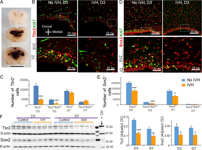
Figure 3.
Occurrence of IVH reduced Sox2 and Tbr2 in preterm rabbits. (A) Coronal brain slice from the frontoparietal lobe of E28.5 rabbit kits which show slit like ventricles in kits without IVH (upper panel) and moderate to severe IVH resulting in fusion of the lateral ventricles (middle and lower panel). Scale bar, 1 cm. (B–E) Representative immunofluorescence of cryosections from E28.5 rabbit kits with and without IVH at D3 (as indicated) labeled with ki67 and Tbr2/Sox2 specific antibodies. Upper panel is low power image and lower panel is high magnification images of the boxed area in the upper panel. Note diminished number of Tbr2+ and Sox2+ cells in rabbits with IVH relative to controls without IVH. The bar charts are mean ± s.e.m. (n = 5 each). The total and cycling Tbr2+ cells were reduced in rabbits with IVH compared with glycerol controls without IVH at D3, not at D7. All Sox2+ cells were reduced in rabbits with IVH compared with glycerol controls without IVH at both D3 and D7. Cycling Sox2+ cells were reduced in rabbits with IVH at D3, not at D7. (F) Representative Western blot analyses for Tbr2 and Sox2 on brain homogenates of preterm rabbits with and without IVH at D3 and D7. The bar charts are mean ± s.e.m. (n = 5 each). Values were normalized to β actin levels. Both Sox2 and Tbr2 levels were reduced in kits with IVH relative to controls without IVH at D3 and D7. ***P < 0.001, **P < 0.01, *P < 0.05 indicate comparison between rabbits with and without IVH at D3 and D7. Scale bar as indicated.

