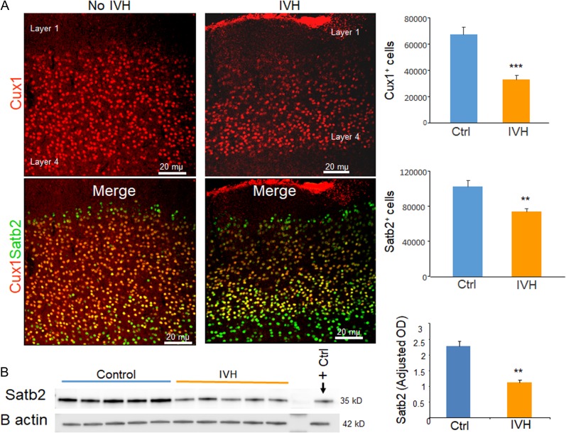
Figure 4.
IVH reduced number of neurons in the upper cortical layer. (A) Representative immunofluorescence of cryosections from preterm rabbits with and without IVH at D14 (as indicated) labeled with Cux1 and Satb2 specific antibodies. Upper panel is Cux1 and lower panel is merge image of Cux1 and Satb2 form upper cortical layer as indicated. Note reduced number of Cux1+ and Satb2+ neurons in rabbits with IVH compared with controls without IVH. The bar charts are mean ± s.e.m. (n = 5 each). Stereological quantification revealed that both Cux1+ and Satb2+ neurons were reduced in rabbits with IVH compared with controls. (B) Representative Western blot analyses for Satb2 on brain homogenates of preterm rabbits with and without IVH at D14. The bar charts are mean ± s.e.m. (n = 5 each). Values were normalized to β actin levels. Satb2 levels were reduced in rabbits with IVH relative to controls. **P < 0.01, ***P < 0.001 indicate comparison between infants with and without IVH.

