Figure 6.
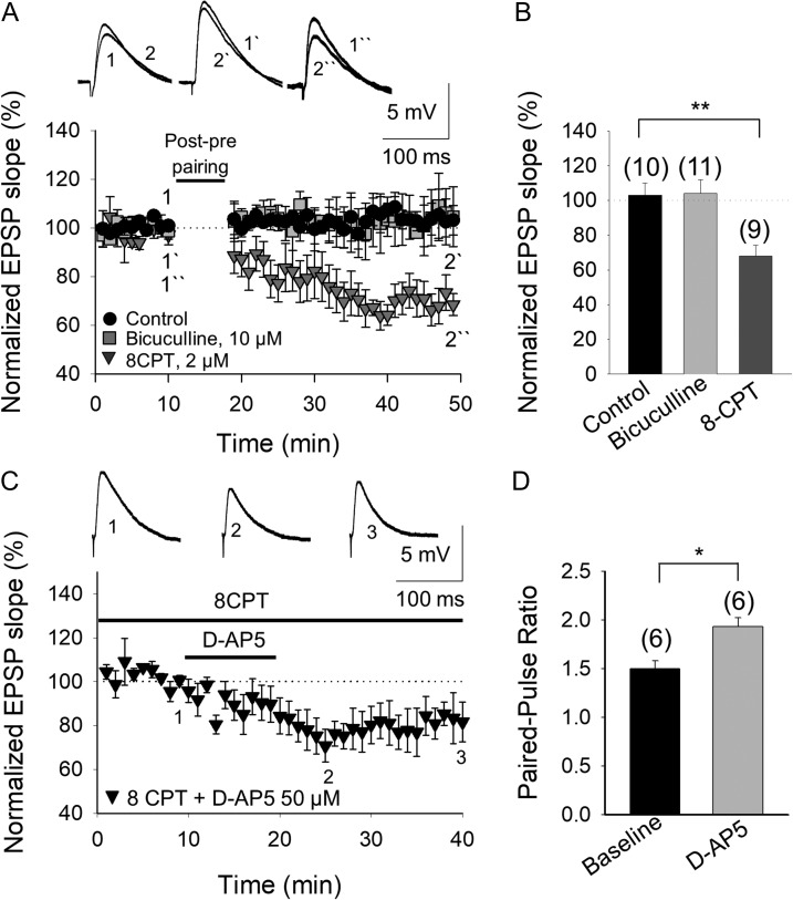
The developmental loss of t-LTD involves an increase in the inhibition mediated by adenosine A1 type receptor activation. (A) The loss of t-LTD is due to the activation of A1Rs and not to the activation of GABAA receptors. The t-LTD lost at P22–P30 was not recovered in the presence of bicuculline (grey squares), whereas the lost t-LTD is completely recovered in the presence of the A1R antagonist 8-CPT (2 μM, grey triangles). The insets show the EPSP before (1, 1´, 1´´) and after (2, 2´, 2´´) post–pre pairing in control conditions (black circles), in the presence of bicuculline or 8-CPT. (B) Summary of the results. (C) The tonic activation of pre-NMDARs that is lost at P22–P30 is completely recovered when A1Rs are antagonized. In the presence of 8-CPT and with the postsynaptic neuron loaded with MK-801, D-AP5 induces a reversible decrease in the EPSP slope. Traces show the EPSP before (1), during (2), and after (3) exposure to D-AP5. (D) The paired-pulse ratio increases in the presence of D-AP5. The error bars indicate the S.E.M. and the number of slices is shown in parentheses: *P < 0.05; **P < 0.01, unpaired Student’s t-test.

