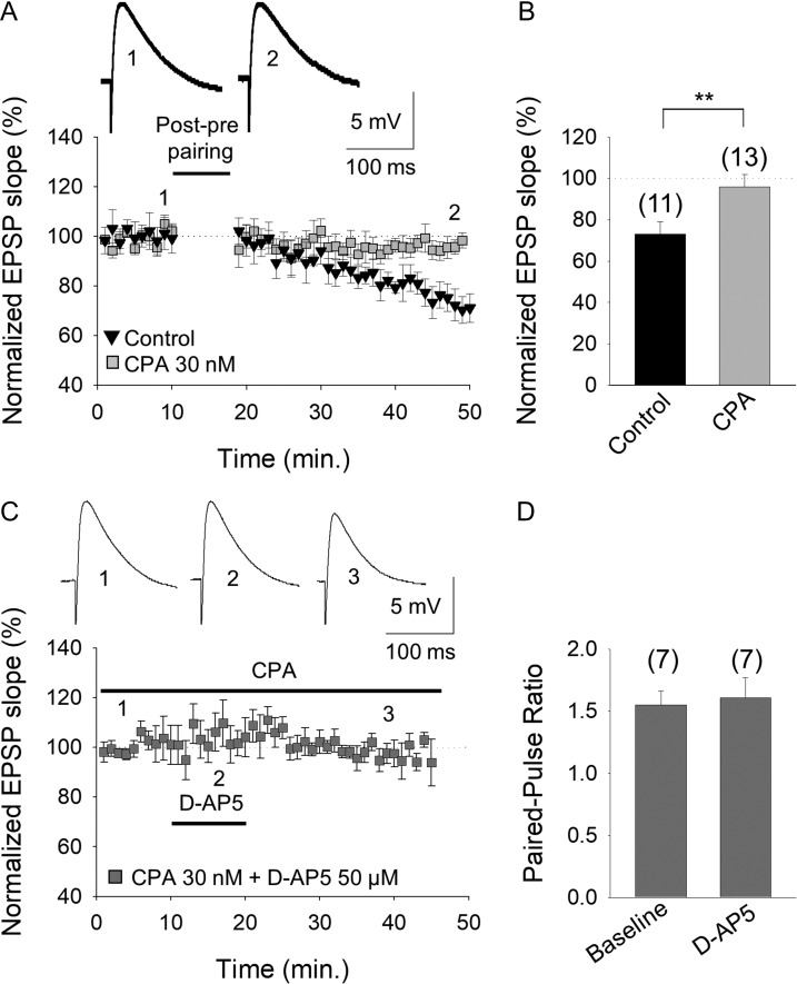
Figure 7.
An increase in A1R mediated-inhibition closes the window of plasticity for t-LTD at P13–P21. (A) Activation of A1Rs at P13–P21 by the agonist CPA prevented the induction of t-LTD. The evoked EPSP slopes monitored in control slices (black triangles) and in slices treated with the A1R agonist CPA (grey squares) following a post–pre pairing are shown. The traces show the EPSP before (1, 1´) and 30 min after (2, 2´) pairing in control slices, and in slices treated with CPA. (B) Summary of the results. (C) Tonic pre-NMDAR activation at P13–P21 is lost in the presence of the A1R agonist CPA. D-AP5 did not affect the evoked EPSP slope in the presence of CPA. The traces show the EPSP before 1), during 2), and after 3) exposure to D-AP5. (D) The paired-pulse ratio was not affected by D-AP5 in the presence of CPA (with the postsynaptic cell loaded with MK-801). The error bars represent the S.E.M. and the number of slices is shown in parentheses: **P < 0.01, unpaired Student’s t-test.

