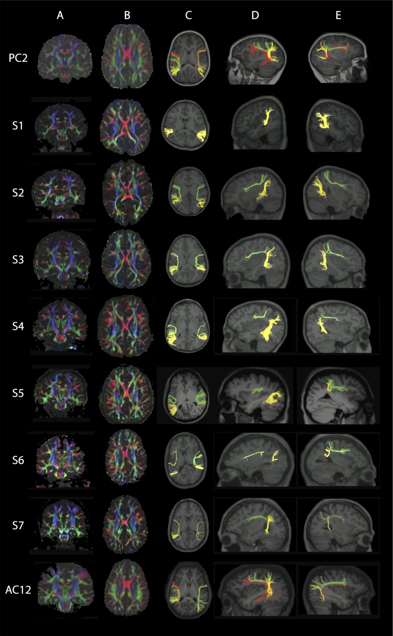
Figure 5.
Dorsal Language Network (DLN). Each row represents images for a single individual with row 1: pediatric control (PC2); rows 2–5: pediatric subjects S1–S4; rows 6–8: adult subjects S5–S7; row 9: adult control (AC12). Column A: Coronal view of color FA maps at the level of the body of the CC (typical radiological convention with left on the right and anterior at top of image). Column B: Axial view of FA color maps at the level of the body of the CC. Note the absence of the DLN.long green region and smaller size of the DLN.ant green region in S1–S7 compared with controls. Color scheme represents the dominant direction of the principal eigenvector in each voxel; as per standard RGB convention for fiber direction, red corresponds to right/left, green corresponds to anterior posterior, and blue corresponds to superior/inferior. The pseudocolored fibers are overlaid on each individual’s structural MPRAGE image, presented in standard radiological convention in columns C and D, while in column E anterior is to the right. Column C: Axial view showing the reconstructed segments of the DLN bilaterally; anterior segment (DLN.ant; green); long segment (DLN.long; red); posterior segment (DLN.post; yellow). Column D: Sagittal view of the left hemisphere shows reconstructed segments of the left DLN. Column E: Sagittal view of the right hemisphere shows reconstructed segments of the right DLN. Note that in all subjects the DLN.long could not be reconstructed. See Supplementary Fig. 5A,B for pediatric and adult controls, respectively.

